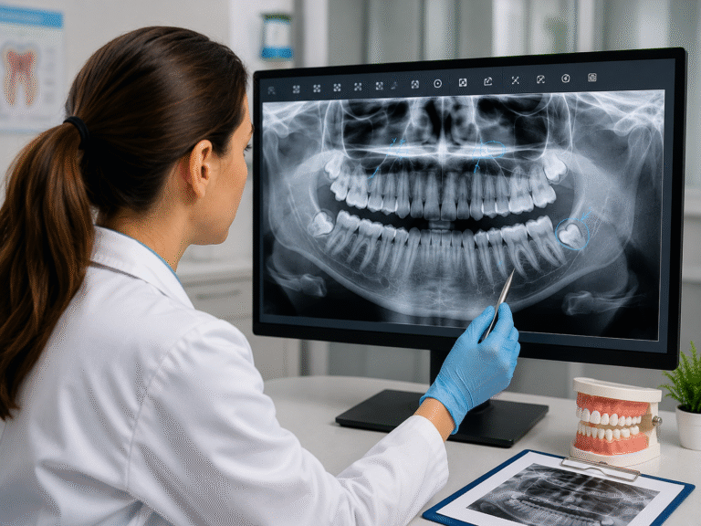
Dental radiographs, or dental X-rays, show what a visual exam cannot, including decay between teeth, infection near the roots, and changes in the surrounding bone. Learning a few basics helps you understand your own results and follow along at your next dental visit.
Interpreting dental X-rays mostly comes down to reading light and dark. Dense parts like enamel and metal fillings look bright, while decay, infection, and bone loss look like darker shadows. This guide covers that light-and-dark rule, a simple reading routine, what cavities look like, and how healthy and unhealthy teeth compare.
Radiopaque vs Radiolucent: The Basics
Every X-ray works on one contrast. Radiopaque (light) areas are dense, such as enamel, crowns, and fillings. Radiolucent (dark) areas are softer or damaged, such as decay and infection. According to the Cleveland Clinic, dental X-rays reveal structures hidden during a normal exam, like the jawbone, roots, and sinuses. Gray tones usually show dentin and the pulp inside each tooth. 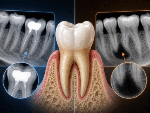
Most clinics now use digital radiography, which gives instant images at a lower dose than older film. You may also see different views, such as bitewing X-rays for decay between the back teeth, periapical X-rays for a full tooth and root, and panoramic X-rays for the whole mouth.
How To Read Dental Radiographs?
To read dental radiographs, check each tooth in the same order from crown to root and compare every structure against healthy enamel. Healthy enamel looks bright and even, while decay, infection, and bone loss look darker. A simple routine helps:
- Find the teeth and any existing fillings, crowns, or implants, which appear bright white.
- Scan the enamel and dentin for dark shadows or notches that can mean decay.
- Check the root tips, where a dark, rounded area may signal an abscess.
- Look at the bone level, since low or uneven bone can point to gum disease.
- Compare both sides, as matching teeth make anything unusual stand out.
Using the same steps each time makes interpreting dental X-rays more reliable and harder to get wrong.
What Do Cavities Look Like on X-Ray?
On an X-ray, a cavity (or dental caries) looks like a dark shadow where a solid tooth should be. Because decay is less dense than enamel, a tooth decay X-ray shows it as a darker area, often well before it causes any pain.
Cavities usually appear in three spots:
- Between teeth: small dark triangles, often first seen on a bitewing tooth decay X-ray.
- On chewing surfaces: darker pits under the enamel.
- Around old fillings: dark margins next to a bright restoration.
As a general rule, the larger and darker the shadow, the deeper the decay tends to be. Catching these areas early often means a smaller filling and less treatment later.
Healthy vs Unhealthy Teeth X-Ray
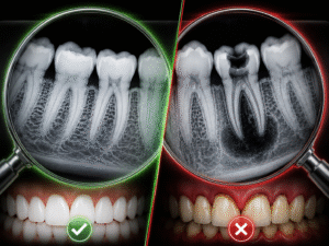
A healthy vs unhealthy teeth X-ray is easiest to judge side by side:
| Feature | Healthy | Unhealthy |
| Enamel | Bright, solid white outline | Dark spots or breaks in the outline |
| Between teeth | Solid contact points | Dark triangles that signal decay |
| Root tip | Even surrounding bone | Dark area, a possible abscess |
| Bone level | High, near the crown | Receded or uneven, showing bone loss |
| Old fillings | Smooth, sealed edges | Dark shadows at the margins |
Common Findings on Dental X-Rays
Beyond reading a tooth decay X-ray, dentists also watch for bone loss from periodontal disease, dark spots near the roots that suggest infection, hardened tartar along the roots, and impacted teeth such as wisdom teeth sitting below the gumline. Each of these shows up as a change in brightness, which is why a careful, consistent read matters.
Conclusion
Once you know that bright means dense and dark often means a problem, dental radiographs become much easier to follow. Still, reading them yourself is only a starting point. As the American Dental Association notes, a diagnosis cannot come from X-rays alone, so always confirm anything unusual with your dentist.
FAQs
Do Cavities Show Up on X-Rays?
Yes. Cavities appear as dark shadows where the tooth has lost density, and X-rays often catch decay between teeth or under fillings that the eye can miss.
How Often Should I Get Dental X-Rays?
It depends on your age and decay risk. Many adults have bitewing X-rays every one to two years, but your dentist sets the right schedule for you.
Are Dental X-Rays Safe?
Yes. Modern digital X-rays use very low radiation, and dentists follow the ALARA principle to keep exposure as low as reasonably achievable.
Can an X-Ray Show Gum Disease?
Yes. Because X-rays reveal the bone supporting your teeth, they can show the bone loss linked to gum disease, sometimes before other symptoms appear.